INTRODUCTION
Fluoxetine, one of the selective serotonin reuptake inhibitors, shows a strong antidepressive effect and is widely used to augment the actions of serotonin (5-HT) in the nervous system.1,2 However, like other antidepressants, the clinical effects of fluoxetine do not begin to appear until a few weeks after the patient has begun taking the drug.3
Previous studies have shown that chronic treatment with antidepressants, including fluoxetine and tianeptine, regulates the expression of genes related to intracellular signal transduction pathways, including pathways involving cyclic adenosine monophosphate (cAMP) responsive element binding protein (CREB), brain-derived neurotropic factor (BDNF), mitogen-activated protein kinase and cell survival, indicating that antidepressants may keep nerve cells from being damaged and also help them regenerate.4-7 Nibuya et al.4 demonstrated that chronic treatment with antidepressants, especially imipramine and fluoxetine, increased CREB and BDNF expression mRNA in rat hippocampus, whereas Reagen et al.6 found that tianeptine increased BDNF protein levels, not in the rat hippocampus (as expected), but in the rat amygdala. This result demonstrated that the brain regions that are targeted by antidepressants can differ. Recently, chronic treatment (8 weeks) of fluoxetine and amitriptyline was shown to increase cyclic guanosine monophosphate but not cAMP in rat hippocampus, suggesting that sustained antidepressant treatment may alter cyclic mononucleotide messenger systems.7 However, it is still not fully known how the expression of genes related to the cellular mechanisms responsible for the therapeutic effect of antidepressants changes during treatment.
In a previous study, we identified genes that may be involved in the therapeutic response of fluoxetine using microarray technology, which can simultaneously monitor the expression levels of thousands of genes.8 In our study using rat basophilic leukemia (RBL-2H3) cells, fluoxetine mediated the up- and down-regulation of several genes, including 14-3-3 eta, which was down-regulated at 24 h and 36 h and then up-regulated at 48 h. Even though our previous research described a new method for analyzing the expression pattern of several genes that are simultaneously regulated by fluoxetine, there were a few limitations (such as the use of cells that did not originate in the brain and the lack of a chronic treatment paradigm for fluoxetine).
The rat C6 glioma cells used in the present study are suitable for studying the effects of antidepressants in vitro because the cells lacking presynaptic input exhibit directly monoamine receptor downregulation through postsynaptic action of the antidepressants.9 In addition, C6 glioma cells have N-methyl-D-aspartate (NMDA) and dopamine D2 receptors as previously mentioned.10,11 Previous studies have observed 5-HT receptor-mediated effects using C6 glioma cells.12,13 Therefore, we can observe a direct postsynaptic effect as well as the biological functions mediated by NMDA, D2 and 5-HT receptors in the cells after fluoxetine treatment. In the present study, we analyzed the genes that were up- and down-regulated by acute (24 h) and chronic (72 h) treatment with fluoxetine in the cells using cDNA microarray. We then confirmed the changes in Gs╬▒, G╬▒i2, and Bcl-xL genes, which were seen in the microarray data, using quantitative RT-PCR (qRT-PCR). In addition, we measured the expression level of Bcl-xL protein after fluoxetine treatment using western blot.
METHODS
Materials
The rat C6 glioma cells (KCLB No;10107) were purchased from KTCC (Seoul, Korea), a rat brain 10K cDNA chip was obtained from Gaiagene (Seoul, Korea) and iQ SYBR green supermix was purchased from Bio-Rad (Hercules, CA, USA). BCL-xL antibody was purchased from Cell Signaling Technology (Beverly, MA, USA) and ╬▓-actin was obtained from Santa Cruz Biotechnology (Santa Cruz, CA, USA). Fluoxetine was provided by Lilly Korea, Ltd. (Seoul, Korea).
Cell culture and fluoxetine treatment
C6 cells were grown overnight in Dulbecco's modified eagle medium containing L-glutamate, 4.5 g/L of glucose, 10% fetal bovine serum, 100 units of penicillin, and 100 ┬Ąg/mL of streptomycin at 37Ōäā in humidified 5% CO2. As a control, the cells were fed with fresh media and cultured for 24 h and 72 h. For the fluoxetine treatment group, the cells were fed with fresh media containing 10 ┬ĄM of fluoxetine and cultured for 24 h and 72 h, as was done in a previous study.14
Microarray analysis
cDNA microarray analysis was performed using a chip containing 2,500 ESTs and 2,500 known genes. The total RNA from the control and fluoxetine-treated cells was isolated using TRIzol┬« Reagent (Invitrogen, Carlsbad, CA, USA) according to the manufacturer's instructions. Fluorescent (Cy3, Cy5)-labeled cDNA probes were made from 20 ┬Ąg of total RNA from both the control cells and the fluoxetine-treated cells, using an amino-allyl cDNA labeling kit (Ambion, Austin, TX, USA). For each experiment, at least four replicates were performed, and two of these replicates were repeated with the fluorophores reversed to eliminate false-positive results. Cy3- and Cy5-labeled probes were mixed with water, 8 ┬Ąg of poly (dA) (Pharmacia), 4 ┬Ąg of Escherichia coli tRNA (Sigma-Aldrich, Saint Louis, MO, USA), 10 ┬Ąg of mouse Cot1 DNA (Invitrogen, Carlsbad, CA, USA) and 60 ┬ĄL of 2X EasyHyb buffer (U-vision Biotech Inc., Taipei, Taiwan) to a final volume of 120 ┬ĄL. The resulting mixture was incubated at 95Ōäā for 5 min. Probes were then applied to the array for hybridization at 52Ōäā for 8 h, using a GeneTAG automatic hybridizer (Genomic Solutions, Boston, MA, USA). After hybridization, the slides were washed first in 2├ŚSSC and 0.2% SDS at 52Ōäā for 5 min, then with 0.1├ŚSSC and 0.2% SDS at 52Ōäā for 5 min and finally with 0.1├ŚSSC for 5 min at room temperature. The slides were dried by spinning for 3 min. The dried slides were scanned with a ScanArray 5,000 fluorescence reader, and QuantArray image acquisition software (PerkinElmer Life Sciences) was used to quantify the signal and background intensity for each target element. The ratio of the two corrected signal intensities was calculated and used as the differential expression ratio (DE) for each specific gene in the two mRNA samples. The raw data of the repeated experiments were converted to log ratios. Negative control spots, small signal-to-noise ratio spots that were less than 2 and spots whose average sensitivity was less than 7.5 were removed, and the data were standardized by Lowess smoothing. Four repeated experiments were standardized by the significant analysis of microarray (SAM; t-statistics) method. Genes differentially expressed by fluoxetine were linkage-analyzed using Cluster 3.0 (open source clustering software) and visualized using JavaTreeview-1.1.3 software (Human Genome Center, Institute of Medical Science, Tokyo, Japan).
qRT-PCR
Total RNA from fluoxetine-treated and control cells was isolated using TRIzol┬« Reagent and reverse transcribed to cDNA in order to confirm the expression patterns of the genes Gs╬▒, G╬▒i2, and Bcl-xL which were differentially expressed by fluoxetine treatment on cDNA microarray result. For qRT-PCR, 12.5 ┬ĄL of SYBR┬« Premix Ex TaqTM II (Takara BIO, Shiga, Japan), 0.5 ┬ĄL of ROX, 5 pmol of forward primer, 5 pmol of reverse primer and 0.5 ┬ĄL of cDNA were mixed with water to a final volume of 25 ┬ĄL. The mixture was amplified for 40 cycles with an ABI 7500 Real-Time PCR System (Applied Biosystems, Inc., Foster City, CA) with an initial melt at 95Ōäā for ten min followed by 40 cycles of 95Ōäā for 15 sec and 60Ōäā for one min. The cycle number at which a statistically significant increase in each gene was first detected (threshold cycle, Ct) was then normalized to the Ct for ╬▓-actin, which was used as an internal control. The relative expression differences between the control and fluoxetine-treated cells were calculated using the 2-╬ö╬öCT method.15 The primers used to amplify the described genes were listed in Table 1. Primers were designed with Genamics Expression software (Genamics, Hamilton, New Zealand).
Western blot analysis
The fluoxetine-treated cells were cultured in media containing 10 ┬ĄM of fluoxetine for 6 h, 24 h, 72 h or 96 h. The cells were then washed in phosphate-buffered saline and harvested. Total protein was extracted from the cells using Pro-PrepTM (iNtRON Biotechnology, Korea) according to the manufacturer's instructions. The protein was quantified using the Bradford method (Bio-Rad Protein Assay Kit; Bio-Rad, Hercules, CA, USA). The protein was separated on 12% polyacrylamide gel through sodium dodecyl sulfate-polyacrylamide gel electrophoresis and electrotransferred onto a nitrocellulose membrane (Bio-Rad, Hercules, CA, USA). After blocking with 5% non-fat dry milk in TBST (20 mM Tris-HCl, pH 7.6; 500 mM NaCl, 0.1% Tween 20), the membrane was incubated overnight at 4Ōäā with the polyclonal rabbit anti-Bcl-xL antibody (1 : 1,000), followed by incubation with HRP-conjugated anti-mouse IgG (diluted 1 : 1,000) for 60 min. After washing in TBST, the reactions were detected using the enhanced chemiluminescence detection system (Amersham Biosciences, Sunnyvale, CA, USA). The concentration of Bcl-xL exposed on X-ray film was quantified with QuantityOne software (Bio-Rad, USA).
Statistical analysis
Data were expressed as the mean┬▒standard error. A nonlinear-fitting program (OriginLab Corp., Northampton, MA, USA) was used. Paired and unpaired Student's t-tests were used as appropriate to evaluate the statistical significance of differences between the two groups (fluoxetine-treated and control groups). Values of p<0.05 were considered to indicate statistical significance.
RESULTS
Differentially expressed genes in fluoxetine-treated C6 cells
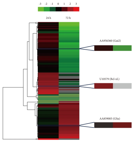
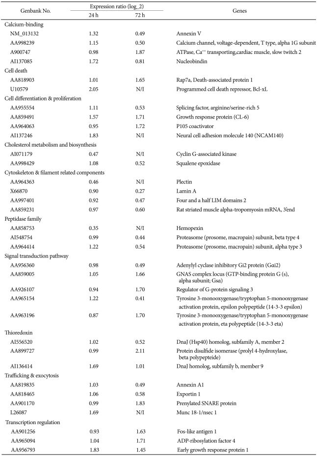
The gene expression profile of control and 10 ┬ĄM fluoxetine-treated cells after 24 h and 72 h was analyzed with a chip containing 2,500 ESTs and 2,500 known genes. After mining biological data using log_2 (a fold change cut-off of 1.5), a hierarchical dendrogram of 169 genes was completed by clustering differentially expressed genes at 24 h and 72 h after fluoxetine treatment (Figure 1). Of the 2,500 known genes in the chip, 7 genes were down-regulated more than 1.4-fold in cells treated with fluoxetine for 24 h compared to the control cells, and 77 of 2,500 known genes were down-regulated after 72 h of fluoxetine treatment. Meanwhile, 33 genes were up-regulated more than 1.4-fold in fluoxetine-treated cells after 24 h compared to control cells, and 53 genes were up-regulated more than 1.4-fold in fluoxetine-treated cells after 72 h of treatment.
Table 2 shows a summarized list of time-dependently expressed genes between the control and fluoxetine-treated C6 cells. Of the down-regulated genes in fluoxetine-treated cells at 24 h, cyclin G-associated kinase, which assists heat shock cognate 70 in uncoating clathrin-coated vesicles16,17 decreased in expression by two-fold. In the fluoxetine-treated cells at 24 h, Bcl-xL and neural cell adhesion molecule 140 (NCAM140) were up-regulated. Bcl-xL (one of the Bcl-2 family members) is widely expressed in the adult brain, despite the relative lack of physiological apoptosis.18
Annexin V and G╬▒i2 were down-regulated in fluoxetine-treated cells at 72 h, whereas 14-3-3 eta and Gs╬▒ were up-regulated. Compared to the expression changes at 24 h, more genes showed a change in expression at 72 h.
Fluoxetine-induced changes in Gs╬▒, G╬▒i2, and Bcl-xL expression
To confirm the result of the microarray analysis, we performed qRT-PCR on RNA isolated from control and fluoxetine-treated cells. Gs╬▒ mRNA expression was 1.03┬▒0.12-fold greater at 24 h and 2.63┬▒0.24-fold greater at 72 h in fluoxetine-treated cells compared to the control cells as observed in our previous study (Figure 2A).15 On the other hand, G╬▒i2 expression was 0.86┬▒0.06-fold at 24 h and 0.72┬▒0.05-fold at 72 h compared to control as observed in our previous study (Figure 2B).15 Gene expression of Gs╬▒ and G╬▒i2 at 24 h was not significantly different from control levels. However, at 72 h, gene expression of Gs╬▒ and G╬▒i2 was significantly different from expression in the control cells. Chronic treatment of fluoxetine induced up-regulation of Gs╬▒ and down-regulation of Gi╬▒2, which was also found in a previous study.19 Bcl-xL mRNA significantly increased by 1.36┬▒0.13-fold at 24 h and by 1.65┬▒0.13-fold at 72 h in fluoxetine-treated cells as compared to control cells (Figure 2C). The expression in all three of these genes changed more after chronic treatment than after acute treatment.
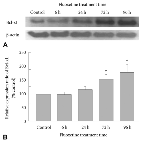
Effect of fluoxetine on Bcl-xL protein expression
After treating C6 cells with 10 ┬ĄM fluoxetine and culturing them for 6 h, 24 h, 72 h or 96 h, we observed the expression pattern of Bcl-xL protein. Expression gradually increased as treatment time progressed (Figure 3A). The increase of Bcl-xL expression at 6 h and 24 h was not significantly different in fluoxetine-treated cells compared to the control cells (Figure 3B). However, at 72 h and 96 h, there was a significant increase in Bcl-xL expression compared to control. Therefore, fluoxetine increased the expression of the Bcl-xL protein over time.
DISCUSSION
Over the past few decades, theories on depression have suggested that atrophy of neurons from stress, apoptosis or depression may result from an impairment of the intracellular signaling pathway and a failure of neurons to appropriately adapt.20,21 Studies on antidepressants and depression have mainly focused on the molecular and cellular antidepressant-induced changes that lead to neurogenesis and restored synaptic activity.
We analyzed the genes that were up- and down-regulated as a result of acute and chronic treatment with fluoxetine in C6 cells and detected expression changes in genes that engage in the intracellular signal transduction cascade, including the following: Gs╬▒ and G╬▒i2; neurite outgrowth, including NCAM140; and cell survival, such as annexin V and Bcl-xL. Depressed patients exhibit dysfunction of the cAMP signaling pathway, namely a decrease in adenylyl cyclases, the enzymes that generate intracellular cAMP.22 This dysfunction occurs in the cerebral cortices of patients with depression. Adenylyl cyclase is known to bind with the stimulatory G protein (Gs) and subsequently increase cAMP and cAMP-dependent protein kinase.23 In the present study, we observed that chronic treatment with fluoxetine induced expression of Gs╬▒ (the stimulatory ╬▒-subunit of the Gs protein) mRNA more than 2.5-fold compared to untreated control cells, a finding that was similar to previous studies.10,24 Toki et al.24 demonstrated that chronic treatment of C6 glioma cells with fluoxetine induced a 2-fold increase of Gs╬▒ in the plasma membrane compared to control cells and also promoted the binding of Gs╬▒ and adenylyl cyclase. Chen and Rasenick10 reported that chronic treatment with desipramine, one of the tricyclic antidepressants, enhanced the binding of Gs╬▒ and adenylyl cyclase by 1.7-fold compared to control. In the present study, G╬▒i2 (an antagonist of Gs╬▒) mRNA expression was seldom different in cells with acute treatment of fluoxetine compared to control cells, but chronic treatment decreased its expression by 2-fold in the fluoxetine-treated cells. In a study by Emamghoreishi et al.,19 chronic treatment with desipramine induced a 10% decrease of G╬▒i2 expression in rat cerebral cortex, but the decrease was not significantly different from expression in the control group. When considering the previous studies10,19,24 along with our work, chronic treatment with antidepressants (specifically fluoxetine) can clearly lead to an increase of Gs╬▒ and a decrease of G╬▒i2, suggesting that fluoxetine may activate the cAMP signaling pathway. This activation induces genes related to neuronal survival and synaptic remodeling.
NCAM, which has three major isoforms (NCAM180, 140 and 120), regulates structural and synaptic plasticity in the brain.25,26 In particular, NCAM140 promotes neurite outgrowth.27 A recent report showed that NCAM140 mRNA is decreased in the peripheral blood cells of patients with bipolar disorder, indicating that this change may be associated with the pathophysiology of the disease.28 Interestingly, we found that NCAM140 mRNA expression was increased after acute fluoxetine treatment. We hypothesize that fluoxetine may promote neurite outgrowth of damaged neural cells in the brains of depressed patients through up-regulation of NCAM140.
Annexin V is a Ca2+-dependent protein that combines with phospholipids phosphatidylserine (PS) after the translocation of PS from the inside to the outside of the plasma membrane. Annexin V expression increases in the early stages of cell death.29 In the present study, we observed that chronic treatment with fluoxetine led to a 2-fold down-regulation of annexin V compared to control cells.
In the present study, Bcl-xL expression (both mRNA and protein) in the C6 cells was up-regulated by both acute and chronic treatment with fluoxetine, revealing a tendency for the expression level of Bcl-xL to rise as the duration of fluoxetine treatment grew longer. As already mentioned, neurotrophic factors (such as CREB and BDNF) are regulated by stress and antidepressants through cell survival pathways that also control the Bcl-2 family proteins.30,31 Of the Bcl-2 family, Bcl-2 and Bcl-xL are localized to the mitochondrial outer membrane, act as antiapoptotic proteins in intracellular apoptotic signal transduction32 and are up-regulated by treatment with antidepressants.5,33 Kosten et al.33 used an animal model of depression to show that both repeated stress and the chronic administration of antidepressants (such as fluoxetine and reboxetine) differentially regulated Bcl-xL mRNA expression in the rat hippocampus. They reported that reboxetine increased Bcl-xL mRNA in the hippocampus but that fluoxetine did not have an effect on Bcl-xL. On the other hand, Chiou et al.5 observed that Bcl-xL and Bcl-2 mRNA expression was up-regulated by chronic treatment with fluoxetine in lipopolysaccharide (LPS)-treated neural stem cells (NSCs) derived from the hippocampus; this up-regulation repressed LPS-induced apoptosis in NSCs. Based on the results of the present study as well as previous studies,5,33 we suggest that when fluoxetine regulates the expression of antiapoptotic agents (Bcl-2 and Bcl-xL), the effects are different in vivo and in vitro; for example, Bcl-2 is up-regulated by fluoxetine in vivo, whereas Bcl-xL is mainly up-regulated in vitro. Furthermore, the increase that we see in Bcl-xL protein levels (as well as in mRNA levels) induced by chronic treatment with fluoxetine clearly indicates that fluoxetine inhibits apoptosis through the Bcl-xL-related antiapoptotic pathway. This result is unlike previous studies, which only demonstrated an increase in Bcl-xL mRNA.5,33
In conclusion, our data not only confirmed that chronic treatment of fluoxetine induces Gs╬▒ and repressed G╬▒i2 expression but also demonstrated that expression of Bcl-xL (both mRNA and protein) is increased by chronic treatment with fluoxetine in C6 cells. These results suggest that these genes may play a critical role in delaying the clinical effects of antidepressants. However, additional studies are necessary to further analyze the other genes found in the microarray and their roles in the effects of antidepressants.