Psychiatry Investig Search

CLOSE


Psychiatry Investig > Volume 17(6); 2020 > Article
Lee, Lee, Byun, Yi, Jung, Park, and Lee: Comparison of Amyloid Positivity Rate and Accumulation Pattern between Amnestic and Non-Amnestic Type Mild Cognitive Impairment

Abstract

Objective

We aimed to compare cerebral beta-amyloid protein (Aβ) positivity rate and amyloid accumulation pattern on amyloid positron emission tomography (PET) between mild cognitive impairment (MCI) subtypes, i.e. amnestic mild cognitive impairment (aMCI) and non-amnestic mild cognitive impairment (naMCI).

Methods

The study participants were 34 naMCI patients and age-, sex- and education-matched 68 aMCI patients (1:2 ratio) who visited the Dementia and Age-Associated Cognitive Decline Clinic of the Seoul National University Hospital. All participants received comprehensive clinical and neuropsychological assessments and [18F] florbetaben PET.

Results

Aβ positivity rate of naMCI group (26.5%) was significantly lower than that of aMCI group (64.7%). Among Aβ positive individuals, there was no difference in Aβ accumulation pattern between naMCI and aMCI.

Conclusion

The findings suggest that MCI subtypes based on impaired cognitive domains have a differential association with brain Aβ deposition, a core pathology of AD. Amnestic subtype of MCI are more closely associated with cerebral Aβ deposition compared to non-amnestic subtype. In contrast, the pattern of amyloid deposition does not appear to have any difference between the subtypes.

INTRODUCTION

Mild cognitive impairment (MCI) is a clinical condition between cognitively normal aging and dementia [1]. Based on the presence of memory impairment, MCI is classified into two subtypes, i.e., amnestic MCI (aMCI) and non-amnestic MCI (naMCI) [2,3]. It has been known that the two subtypes of MCI have different clinical characteristics [4,5]. In particular, individuals with aMCI are more likely to progress to Alzheimer’s disease (AD) dementia compared to those with naMCI [6-8].
In regard of in vivo neuropathological substrates underlying each MCI subtypes, however, sufficient information is still not available. Although several amyloid PET studies indicated that the positivity rate of beta-amyloid protein (Aβ) deposition, a core neuropathology of AD, is higher in aMCI than in naMCI [9-12], the sample sizes of naMCI subjects included in previous studies were too small (only 7-18) to make any conclusion. In addition to Aβ positivity rate, one previous study [11] investigated the patterns of cerebral Aβ accumulation in MCI patients suggesting two patterns of accumulation, i.e., A-pattern and B-pattern. In A-pattern, the deposition of cerebral Aβ is mainly observed in the frontal part of the brain. In B-pattern, on the other hand, the deposition is observed evenly throughout the whole brain. While the study reported that 11 out of 36 amyloid-positive aMCI patients had an A-pattern and 1 out of 3 amyloid-positive naMCI patients had an A-pattern [11], further investigation is still needed for larger naMCI sample.
Therefore, we aimed to examine the differences of amyloid positivity rate and accumulation pattern between aMCI and naMCI subtypes in MCI subjects including more naMCI individuals compared to previous studies.

METHODS

Participants

A total of 102 MCI (34 naMCI and age-, sex-, and education matched 68 aMCI) patients were recruited from the pool of older adults who visited the Dementia and Age-Associated Cognitive Decline Clinic of the Seoul National University Hospital (SNUH) to get outpatient clinical services. All participants met the current consensus criteria for MCI: 1) cognitive impairment confirmed by an informant, 2) objective cognitive impairment, 3) preserved global cognitive function, 4) independence in functional activities, and 5) not demented [13,14]. aMCI and naMCI subjects met different criterion 2). For aMCI, the age-, sex-, and education-adjusted z-score for at least one of the four episodic memory tests (i.e., Word List Memory, Word List Recall, Word List Recognition, and Constructional Recall tests) in the Korean version of the Consortium to Establish a Registry for Alzheimer’s disease (CERAD-K) neuropsychological battery [15,16] was below -1.5. In contrast, for naMCI, the age-, sex-, and education-adjusted z-score for all the four episodic memory tests were identical to or above -1.5 and at least one of four non-memory tests (i.e., Boston Naming, Visuospatial Construction, and Sematic Fluency in the CERAD-K neuropsychological battery, and Stroop Color Word test) was below -1.5. All participants had an overall clinical dementia rating (CDR) [17] of 0.5. Exclusion criteria for all subjects were any serious medical or neurological diseases present that could affect mental function, evidence of focal brain lesions on magnetic resonance imaging, the presence of severe behavioral or communication problems that would make a clinical examination difficult, and the absence of a reliable informant. Individuals with minor physical abnormalities (e.g., diabetes with no serious complications, essential hypertension, and mild hearing loss) were included. This study was approved by the Institutional Review Boards of the SNUH (IRB No. 1905-110-1035).

Clinical and neuropsychological assessments

All participants underwent comprehensive clinical and neuropsychological evaluation based on the CERAD-K assessment packet [15,16]. Clinical diagnosis and CDR rating was made through consensus case conference in which more than four psychiatrists with expertise for dementia research participated. The presence or absence of six cerebrovascular risk factors, including stroke, diabetes, dyslipidemia, transient ischemic attack, hypertension, and coronary artery disease, was systematically assessed from the subjects and subjects’ histories provided by the informant as well as a review of pertinent medical records. To calculate an overall measure of cerebrovascular burden, we created a composite score, i.e., the vascular risk score (VRS), which was the sum of the factors present, ranging from 0 to 6 and reported as a percentage [18]. Apolipoprotein E (APOE) genotyping was implemented for subset of participants (n=81).

Assessment for amyloid positivity and accumulation pattern

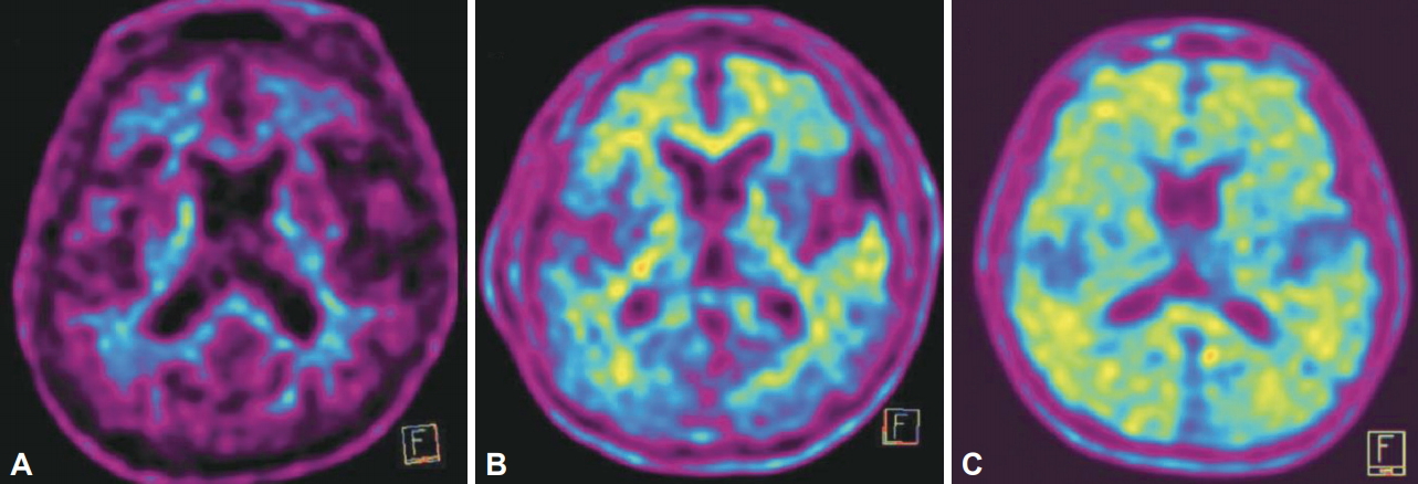
All participant received [18F] florbetaben positron emission tomography (PET) scan. PET scans were performed using the ECAT EXACT47scanner (Siemens-CTI; Knoxville, TN, USA). The participants were classified as either Aβ positive or negative according to the Brain Amyloid Plaque Load (BAPL) score [19]. The BAPL is a three-grade scoring system, which is determined by visual assessment of the degree of Aβ accumulation. If the BAPL score is 2 or 3 points in at least one of the four regions of Interest (ROIs), i.e., the lateral temporal lobes, frontal lobes, posterior cingulate/precuneus and parietal lobes, the patient is classified to Aβ positive. A trained psychiatric resident (SHL) rated the BAPL score for PET scan of every participant. In order to verify the reliability of the rating, the agreement between ratings of the rater (SHL) and clinical ratings provided by nuclear medicine physicians at the SNUH was evaluated by calculating kappa value. More than substantial agreement was observed: Kappa value was 0.729 for aMCI, 1.000 for naMCI, and 0.822 for overall MCI cases. For Aβ positive participants, Aβ accumulation pattern was also determined by visual inspection by the same rater (SHL). The accumulation pattern was classified into A-pattern and B-pattern according to the guideline described in the previous report [11]. A-pattern has predominant amyloid retention in the frontal, anterior cingulate, lateral temporal, and basal ganglia, while B-pattern shows generalized amyloid retention over the entire cerebral cortex (Figure 1).

Statistical analysis

All statistical analyses were conducted using SPSS (version 24, IBM Corp., Armonk, NY, USA). For the comparison between aMCI and naMCI, Student’s t-test or chi-square test was used depending on the type of variables. The threshold for statistical significance was p<0.05 for all the analyses.

RESULTS

Demographic and clinical characteristics

Table 1 shows the demographic and clinical characteristics of the participants by MCI subtype. Since the participants of aMCI and naMCI were matched by age, sex and years of education, there was no significant difference in the demographic factors. The two MCI subtypes also did not differ in regard of age of onset, duration of illness, APOE ε4 carrier state, and VRS. The mean CDR sum of box (SOB) score of aMCI was significantly higher than that of naMCI. As expected, z-scores of four episodic memory tests in the CERAD-K neuropsychological batter were significantly lower in aMCI than in naMCI.

Aβ positivity rate

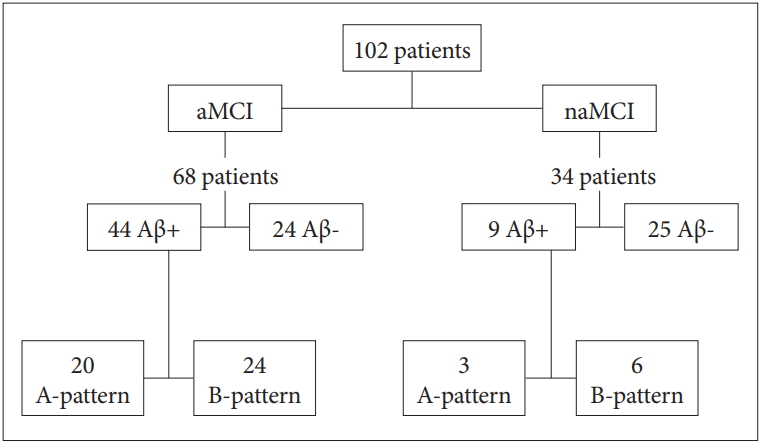
The Aβ positivity rate of aMCI and naMCI was 64.7% and 26.5%, respectively (Table 2, Figure 2). The rate of naMCI was significantly lower than that of aMCI.

Aβ accumulation pattern

There was no significant difference in the proportion of amyloid accumulation patterns between Aβ positive aMCI and Aβ positive naMCI (Table 3, Figure 2).

DISCUSSION

In the present study, cerebral Aβ positivity rate was significantly higher in aMCI compared with naMCI. There was no difference in the proportion of Aβ accumulation patterns between aMCI and naMCI cases with amyloid positivity.
The Aβ positivity rate for aMCI (64.7%) observed in this study was very similar to the rates reported in previous studies, which ranged from 57.9% to 67.9% [9-12]. In regard of the Aβ positivity rate for naMCI, our result obtained from relatively larger sample [9 of 34 (26.5%)] was slightly higher than the rates from two previous studies with smaller sample [3 of 17 (17.6%) [10] and 3 of 16 (18.8%) [11]], while lower than those from other two small studies [6 of 18 (33.3%) [9] and 3 of 7 (42.9%) [12]]. As various factors including demographic variables, study settings, PET ligands used to measure cerebral Aβ, and the definition or threshold for Aβ positivity may influence on the rate, so it is not easy to directly compare it between the studies. Nevertheless, as shown in our study, the Aβ positivity rate of aMCI was higher than that of naMCI across all the studies, consistent with clinical observation that higher proportion of individuals progressed to AD dementia in aMCI than in naMCI [6-8].
We also compared the proportion of Aβ accumulation patterns between aMCI and naMCI and did not find any significant difference. This observation indicates that regional distribution pattern of cerebral Aβ accumulation is not associated with the difference of impaired cognitive domain in MCI. A previous study also reported similar result although the study included only three Aβ positive naMCI subjects [11].
Although previous clinical studies suggested that naMCI individuals are more related with vascular dementia than aMCI ones [6,20], we did not find any difference of VRS between the two subtypes. The lack of significant difference might be attributable to the small sample size or measurement method for vascular burden. When compared for the proportion of subjects who have at least one vascular risk factor, there were significant difference between aMCI and naMCI (70.6% for aMCI vs. 88.2% for naMCI, p=0.048).
Although the present study has a strength in that relatively large number of naMCI patients were included, a couple of limitations should also be mentioned. First, as the MCI subjects were recruited from a dementia clinic in tertiary hospital, there may be some limitation to generalize the results to community population. Second, this study did not evaluate other pathologies frequently found in brain of older adults, such as cerebral tau, alpha synuclein, TDP-43, and vascular pathologies. To get more understandings for the causative neuropathologies underlying MCI subtypes, future studies using other brain imaging modalities or post-mortem brains are necessary.
In conclusion, the current findings suggest that MCI subtypes based on impaired cognitive domains have a differential association with brain Aβ deposition, a core pathology of AD. Amnestic subtype of MCI are more closely associated with cerebral Aβ deposition compared to non-amnestic subtype. In contrast, the pattern of amyloid deposition does not appear to have any difference between the subtypes.

ACKNOWLEDGEMENTS

This study was supported by a grant from Ministry of Science and ICT, Republic of Korea (Grant No. NRF-2014M3C7A1046042) and a grant of the Korea Health Technology R&D Project through the Korea Health Industry Development Institute (KHIDI), funded by the Ministry of Health & Welfare, Republic of Korea (Grant No: HI18C0630 & HI19C0149).

The authors have no potential conflicts of interest to disclose.

Author Contributions

Conceptualization: Dong Young Lee, Jun Ho Lee, Sun Hyung Lee. Data curation: Jun Ho Lee, Sun Hyung Lee. Formal analysis: Sun Hyung Lee, Jun Ho Lee. Funding acquisition: Dong Young Lee. Investigation: Dong Young Lee, Jun Ho Lee, Min Soo Byun, Dahyun Yi, Gijung Jung, Jee Eun Park. Methodology: Dong Young Lee, Jun Ho Lee, Min Soo Byun, Dahyun Yi. Project administration: Dong Young Lee, Jun Ho Lee, Min Soo Byun, Dahyun Yi, Gijung Jung, Jee Eun Park. Resources: Dong Young Lee, Jun Ho Lee, Min Soo Byun. Supervision: Dong Young Lee, Jun Ho Lee. Validation: Dong Young Lee, Jun Ho Lee. Visualization: Sun Hyung Lee. Writing—original draft: Sun Hyung Lee. Writing—review & editing: Dong Young Lee, Jun Ho Lee, Min Soo Byun, Dahyun Yi, Gijung Jung, Jee Eun Park.

Figure 1.
Cerebral beta-amyloid protein (Aβ) accumulation patterns of [18F] florbetaben PET image. A: Aβ negative. B: Predominant Aβ retention in frontal, anterior cingulate, lateral temporal, and basal ganglia (A-pattern). C: Generalized Aβ retention (B-pattern).
pi-2020-0063f1.jpg
Figure 2.
Distribution of cerebral beta-amyloid protein (Aβ) positive and negative cases in amnestic mild cognitive impairment (aMCI) and non-amnestic mild cognitive impairment (naMCI), and distribution of A-Pattern and B-Pattern accumulation in Aβ positive aMCI and naMCI.
pi-2020-0063f2.jpg
Table 1.
Demographic, clinical and neuropsychological characteristics of mild cognitive impairment subtypes
Variables aMCI (N=68) naMCI (N=34) p-value
Demographic and clinical characteristics
 Age (y) 74.6±6.5 74.3±6.9 0.801
 Female, N (%) 58 (85.3) 29 (85.3) 1.000
 Education (y) 9.37±4.97 8.8±4.71 0.597
 Age of onset (y) 71.7±7.5 71.5±7.2 0.910
 Duration of illness (y) 2.99±2.31 2.76±1.58 0.617
 APOE4 carrier, N (%)* 22 (40.0) 7 (26.9) 0.252
 CDR SOB 1.71±0.66 1.43±0.64 0.044
 VRS total 1.31±1.08 1.65±1.10 0.142
Neuropsychological tests (z-score)
 MMSE -1.12±0.98 -0.42±0.85 0.001
 Semantic fluency -0.69±0.96 -0.76±0.84 0.721
 Boston naming -0.11±1.34 -0.26±1.23 0.574
 Word list memory -1.09±0.94 -0.25±0.57 <0.001
 Word list recall -1.70±0.92 -0.23±0.76 <0.001
 Word list recognition -1.36±1.82 -0.15±0.76 <0.001
 Constructional praxis -0.24±1.14 -0.25±1.05 0.971
 Constructional recall -1.25±0.84 -0.32±0.76 <0.001
 Stroop color-word -0.92±1.09 -1.26±0.95 0.115

Data are presented as means±SD for continuous variables and as N (%) for categorical variables with p-values for comparison between aMCI and naMCI. Continuous variables were analyzed through Student’s t-test and categorical variables were analyzed through chi-square test.

* number of participants who underwent APOE genotyping were 81 (aMCI 55, naMCI 26).

aMCI: amnestic mild cognitive impairment, naMCI: non-amnestic mild cognitive impairment, APOE4: apolipoprotein E ε4, CDR-SOB: Clinical Dementia Rating sum of box, VRS: vascular risk score, MMSE: Mini-Mental State Examination

Table 2.
Comparison of Aβ positivity rate between mild cognitive impairment subtypes
aMCI (N=68) naMCI (N=34) p-value
Aβ positive 44 (64.7) 9 (26.5) <0.001
Aβ negative 24 (35.3) 25 (73.5)

Data are presented as N (%). P-value for comparison of aMCI and naMCI with chi-square test. Aβ: beta-amyloid protein, aMCI: amnestic mild cognitive impairment, naMCI: non-amnestic mild cognitive impairment

Table 3.
Comparison of the proportion of Aβ accumulation patterns between Aβ positive aMCI and naMCI
aMCI (N=44) naMCI (N=9) p-value
A-pattern 20 (45.5) 3 (33.3) 0.504
B-pattern 24 (54.5) 6 (66.7)

Data are presented as N (%). P-value for comparison of aMCI and naMCI with chi-square test. Aβ: beta-amyloid protein, aMCI: amnestic mild cognitive impairment, naMCI: non-amnestic mild cognitive impairment

REFERENCES

1. Winblad B, Palmer K, Kivipelto M, Jelic V, Fratiglioni L, Wahlund LO, et al. Mild cognitive impairment-beyond controversies, towards a consensus: report of the International Working Group on Mild Cognitive Impairment. J Intern Med 2004;256:240-246.
crossref pmid
2. Petersen RC, Morris JC. Mild cognitive impairment as a clinical entity and treatment target. Arch Neurol 2005;62:1160-1163.
crossref pmid
3. Gauthier S, Reisberg B, Zaudig M, Petersen RC, Ritchie K, Broich K, et al. Mild cognitive impairment. Lancet 2006;367:1262-1270.
crossref pmid
4. Petersen RC. Mild cognitive impairment. Continuum (Minneap Minn) 2016;22:404-418.
crossref pmid pmc
5. Roberts R, Knopman DS. Classification and epidemiology of MCI. Clin Geriatr Med 2013;29:753-772.
crossref pmid
6. Nordlund A, Rolstad S, Klang O, Edman A, Hansen S, Wallin A. Two-year outcome of MCI subtypes and aetiologies in the Goteborg MCI study. J Neurol Neurosurg Psychiatry 2010;81:541-546.
crossref pmid
7. Mitchell J, Arnold R, Dawson K, Nestor PJ, Hodges J. Outcome in subgroups of mild cognitive impairment (MCI) is highly predictable using a simple algorithm. Neurology 2009;256:1500-1509.
crossref pdf
8. Rasquin S, Lodder J, Visser P, Lousberg R, Verhey F. Predictive accuracy of MCI subtypes for Alzheimer’s disease and vascular dementia in subjects with mild cognitive impairment: a 2-year follow-up study. Dement Geriatr Cogn Disord 2005;19:113-119.
crossref pmid
9. Patricio CM, Gabriela C, Julieta RM, Marcos FS, Federico N, Griselda R, et al. Concordance between 11C-PIB-PET and clinical diagnosis in a memory clinic. Am J Alzheimers Dis Other Demen 2015;30:599-606.
crossref pmid pmc
10. Jiménez-Bonilla JF, Banzo I, De Arcocha-Torres M, Quirce R, MartínezRodríguez I, Lavado-Pérez C, et al. Diagnostic role of 11C-Pittsburgh compound B retention patterns and glucose metabolism by fluorine-18-fluorodeoxyglucose PET/CT in amnestic and non-amnestic mild cognitive impairment patients. Nucl Med Commun 2016;37:1189-1196.
crossref pmid
11. Banzo I, Jimenez-Bonilla JF, Martinez-Rodriguez I, Quirce R, de Arcocha-Torres M, Bravo-Ferrer Z, et al. Patterns of 11C-PIB cerebral retention in mild cognitive impairment patients. Rev Esp Med Nucl Imagen Mol 2016;35:171-174.
crossref pmid
12. Wolk DA, Price JC, Saxton JA, Snitz BE, James JA, Lopez OL, et al. Amyloid imaging in mild cognitive impairment subtypes. Ann Neurol 2009;65:557-568.
crossref pmid pmc
13. Petersen RC. Mild cognitive impairment as a diagnostic entity. J Intern Med 2004;256:183-194.
crossref pmid
14. Petersen RC, Doody R, Kurz A, Mohs RC, Morris JC, Rabins PV, et al. Current concepts in mild cognitive impairment. Arch Neurol 2001;58:1985-1992.
crossref pmid
15. Lee DY, Lee KU, Lee JH, Kim KW, Jhoo JH, Kim SY, et al. A normative study of the CERAD neuropsychological assessment battery in the Korean elderly. J Int Neuropsychol Soc 2004;10:72-81.
crossref pmid
16. Lee JH, Lee KU, Lee DY, Kim KW, Jhoo JH, Kim JH, et al. Development of the Korean version of the Consortium to Establish a Registry for Alzheimer’s Disease Assessment Packet (CERAD-K) clinical and neuropsychological assessment batteries. J Gerontol B Psychol Sci Soc Sci 2002;57:47-53.
crossref
17. Morris JC. The Clinical Dementia Rating (CDR): current version and scoring rules. Neurology 1991;41:1588-1592.
crossref pmid
18. DeCarli C, Mungas D, Harvey D, Reed B, Weiner M, Chui H, et al. Memory impairment, but not cerebrovascular disease, predicts progression of MCI to dementia. Neurology 2004;63:220-227.
crossref pmid pmc
19. Sabri O, Seibyl J, Rowe C, Barthel H. Beta-amyloid imaging with florbetaben. Clin Transl Imag 2015;3:13-26.
crossref pdf
20. Busse A, Hensel A, Gühne U, Angermeyer M, Riedel-Heller S. Mild cognitive impairment: long-term course of four clinical subtypes. Neurology 2006;67:2176-2185.
crossref pmid


ABOUT
AUTHOR INFORMATION
ARTICLE CATEGORY

Browse all articles >

BROWSE ARTICLES
Editorial Office
#522, 27, Seochojungang-ro 24-gil, Seocho-gu, Seoul 06601, Korea
Tel: +82-2-717-0892    E-mail: psychiatryinvest@gmail.com                

Copyright © 2024 by Korean Neuropsychiatric Association.

Developed in M2PI

Close layer
prev next