Psychiatry Investig Search

CLOSE


Psychiatry Investig > Volume 20(12); 2023 > Article
Sun, Wen, Luo, Hu, Ren, Lv, Zhang, Gao, Xuan, Wang, Li, Xiang, and Luan: Positive Association of TEAD1 With Schizophrenia in a Northeast Chinese Han Population

Abstract

Objective

Schizophrenia is a complex and devastating psychiatric disorder with a strong genetic background. However, much uncertainty still exists about the role of genetic susceptibility in the pathophysiology of schizophrenia. TEA domain transcription factor 1 (TEAD1) is a transcription factor associated with neurodevelopment and has modulating effects on various nervous system diseases. In the current study, we performed a case-control association study in a Northeast Chinese Han population to explore the characteristics of pathogenic TEAD1 polymorphisms and potential association with schizophrenia.

Methods

We recruited a total of 721 schizophrenia patients and 1,195 healthy controls in this study. The 9 single nucleotide polymorphisms (SNPs) in the gene region of TEAD1 were selected and genotyped.

Results

The genetic association analyses showed that five SNPs (rs12289262, rs6485989, rs4415740, rs7113256, and rs1866709) were significantly different between schizophrenia patients and healthy controls in allele or/and genotype frequencies. After Bonferroni correction, the association of three SNPs (rs4415740, rs7113256, and rs1866709) with schizophrenia were still evident. Haplotype analysis revealed that two strong linkage disequilibrium blocks (rs6485989-rs4415740-rs7113256 and rs16911710-rs12364619-rs1866709) were globally associated with schizophrenia. Four haplotypes (C-C-C and T-T-T, rs6485989-rs4415740-rs7113256; G-T-A and G-T-G, rs16911710-rs12364619-rs1866709) were significantly different between schizophrenia patients and healthy controls.

Conclusion

The current findings indicated that the human TEAD1 gene has a genetic association with schizophrenia in the Chinese Han population and may act as a susceptibility gene for schizophrenia.

INTRODUCTION

Schizophrenia is a common and severe mental illness with a lifetime risk of around 1% [1]. Although advances in genomics, epidemiology, and neuroscience have led to great progress in our understanding of schizophrenia over the past few decades, its etiology and pathophysiology remain unclear. Like other polygenic genetic diseases, the cause of schizophrenia is complex and multifactorial, with the contribution of many susceptibility genes, epigenetic and environmental factors, as well as random factors [2]. Studies of families, twins, and foster children have all shown that genetic factors are significant in the occurrence of schizophrenia, accounting for about 80% [3]. With the application of the genome-wide association study, more and more susceptible genes of small relation to the risk of schizophrenia have been discovered. Among them, a variety of genes are related to neural development. Neurodevelopmental hypothesis of schizophrenia is a broadly accepted paradigm based on epidemiology, anatomical, and neuroimaging studies, with the view that the symptoms of schizophrenia are the terminal result of developmental problems in the brain resulted from hereditary factors and perinatal environment factors.
TEA domain transcription factor 1 (TEAD1) is a member of the TEAD (TEA/ATTS domain) transcription factor family and mapped to the human chromosome 11p15, a potentially relevant region for schizophrenia [4,5]. TEAD proteins are evolutionarily conserved with an N terminal DNA-binding domain called the TEA domain and a C terminal protein-binding domain which co-activators bind to. TEAD1 can hardly induce transcription of target gene on its own [6]. As is known to all, TEAD1 is a key transcription factor in Hippo signaling pathway, a highly conserved pathway that plays a crucial role in neurodevelopmental stability and nervous system diseases controlled by modulating neuroinflammation, neuronal differentiation, and cell death [7]. Although the transcriptional activity of TEAD1 is traditionally believed to be regulated by the co-activators such as nuclear Yes-associated protein (YAP)/transcriptional coactivator with PDZ-binding motif, several studies suggested that TEAD1 is regulated through Hippo independent mechanisms [8]. Once TEAD1 binds its co-activators, the complexes can regulate the expression of target genes that play an important role in cell growth, proliferation, differentiation, apoptosis, and cell migration. Particularly, TEADs have been direct implicated in development, with expression detected in a wide range of embryonic brains as early as 2-cell stage, especially in forebrain [9]. During cortical development, the expression of TEAD1 increased significantly from the expansion and neurogenesis phase to the gliogenic phase, indicating potential specific functions [10]. TEADs affect the development of different tissues and organs, such as heart [11], muscle [12], neural crest and notochord [13,14], and trophectoderm [15], thus, dysfunction of TEADs can lead to various developmental disease. The knockout of TEAD1 has led to embryo death [11]. Overexpressing a transcriptionally active form of TEAD1 leads to severe dysplasia with reduced neuronal differentiation, increased neural progenitor cells (NPCs), the formation of tumor-like rosettes and injuries of ventricular surface [16]. In addition to development, TEAD1 also play a role in brain tumors generation [17].
In the present study, an independent case-control study was conducted to seek the genetic association of the human TEAD1 gene with schizophrenia in a Northeast Chinese Han population with 721 schizophrenia patients and 1,195 healthy controls.

METHODS

Subjects

We recruited 721 schizophrenia patients and 1,195 healthy controls (Table 1), all unrelated Han Chinese origin, from the Department of Psychiatry, Dalian Seventh People’s Hospital, Northeast China. Diagnoses were made strictly in accordance with the Diagnostic and Statistical Manual of Mental Disorders, Fifth Edition [18], based on the patients’ clinical symptoms, impaired social functioning, course of illness, by two senior psychiatrists. Healthy controls in this study were demographically matched to patients and all had been confirmed by a reliable psychiatrist that they were free of psychiatric disorders. Before any study-related procedures were performed, all participants were aware of the main purpose and methods of the study and provided written informed consent. The study was approved by the Ethics Committee of the Dalian Seventh People’s Hospital, China (ID: 2020-18).

Single nucleotide polymorphism selection

We downloaded information of all single nucleotide polymorphisms (SNPs) within and adjacent to the human TEAD1 gene from the International HapMap project database on db-SNP (http://www.ncbi.nlm.nih.gov/SNP/), and then selected potentially relevant 9 SNPs, of which the minor allele frequencies are all greater than 0.01. The 9 SNPs are evenly distributed in the TEAD1 gene region, and the distance between the adjacent two SNPs is less then 60 kb which is the predicted genetic linkage distance in human. In addition, the selected 9 SNPs covers about 255 kb in length, covering approximately 100% of the total length of the human TEAD1 gene, which is 270 kb.

Genotyping

The venous blood of all subjects was collected to extract genomic DNA by a commercially QIAamp DNA Blood Mini Kit (QIAGEN, Hilden, German). Direct DNA sequencing or polymerase chain reaction (PCR)-restriction fragment length polymorphism analysis was used for genotyping of SNPs and Software Primer Premier 5.0 (Premier, Toronto, Canada) was used to design all PCR primer pairs. PCR products were either separated by agarose gel electrophoresis (2%-3% gel) stained with ethidium bromide after completely digested overnight with 4U of restriction enzyme or sequenced. All the results were independently read by two skilled technicians.

Statistics

All the data were analyzed by SHEsis software (SHEsis Online Version; http://analysis.bio-x.cn/SHEsisMain.htm) [19,20]. and Haploview (Broad Institute, Cambridge, MA, USA; https://www.broadinstitute.org/haploview/haploview). Chi-square test was applied to perform Hardy-Weinberg equilibrium on the whole sample. Statistical differences of allele and genotype frequencies between cases and controls were detected by Pearson χ2 test. D’ value was used to evaluate pairing linkage disequilibrium (LD) between alleles. Odds ratios and their 95% confidence interval were used to evaluate the statistical efficacy of different alleles and haplotypes. The Bonferroni correction was adopted to reduce the class I errors caused by multiple tests, that is, Bonferroni corrected p-value=α/the number of SNP sites. If the p-value of each SNP site is less than Bonferroni’s corrected p-value, it indicates that there is a significant correlation between this site and the disease. Criterion for statistical significance was set at two-tailed p<0.05.

RESULTS

Allele and genotype association of selected TEAD1 SNPs with schizophrenia

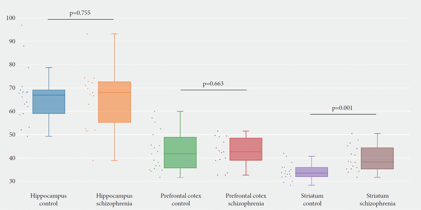
We have checked the expression profile of human TEAD1 gene in the human protein atlas database (https://www.proteinatlas.org/) [21,22], which indicated the TEAD1 gene is broadly expressed in brain tissue with low region specificity (Figure 1A and B). Data from Tabula Muris databases (https://tabulamuris.ds.czbiohub.org/) [23] show that TEAD1 mRNA is highly expressed in the Brain_Non-Myeloid, especially expressed in Bergmann glial cell, brain pericyte, and astrocyte of the cerebral cortex (Figure 1C). However, the data extracted from the SZDB database [24,25] showed that, other than other detected regions, a significant difference of TEAD1 expression in stratum existed between the schizophrenia patients and healthy control subjects (corrected p=0.001) (Figure 2). Therefore, we suspected that the differential expression of TEAD1 in striatum between schizophrenic patients and healthy controls indicates that the human TEAD1 gene may be associated with schizophrenia.
We selected 9 SNPs in the TEAD1 gene region and performed allele and genotype association studies of these SNPs in a case-control sample consisted of 721 schizophrenia patients and 1,195 healthy controls. Detailed information and location of these selected SNPs are shown in Table 2, and none of the genotype distributions of these 9 SNPs in case and control groups deviated from Hardy-Weinberg equilibrium. Of the 9 SNPs, we found remarkable differences in both allele and genotype frequencies of four SNPs between cases and controls: rs6485989-SNP1 (allele: χ2=6.313, p=0.012; genotype: χ2=6.799, p=0.034), rs4415740-SNP2 (allele: χ2=11.564, p=0.001; genotype: χ2=13.024, p=0.002), rs7113256-SNP3 (allele: χ2=11.493, p=0.001; genotype: χ2=12.867, p=0.002), and rs1866709-SNP7 (allele: χ2=8.185, p=0.004; genotype: χ2=8.185, p=0.017) (Table 3). In addition, one SNP only showed significant association in allele frequency: rs12289262-SNP4 (χ2=4.838, p=0.028) (Table 3). After the Bonferroni correction, three SNPs remained significantly associated with schizophrenia in the allele or/and genotype frequencies: SNP2 (allelic p=0.006; genotypic p=0.014), SNP3 (allelic p=0.006; genotypic p=0.015) and SNP7 (allelic p=0.018) (Table 3).

Haplotype analysis of selected TEAD1 SNPs with schizophrenia

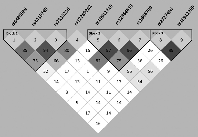
To further explore the association of haplotype structure, the standardized measure D’ value was used to evaluate pairwise linkage disequilibrium of the selected 9 SNPs of TEAD1. When D’ value of two SNPs were between 0.8 and 1.0, there is a strong linkage disequilibrium between them. We found three strong linkage disequilibrium haplotypes, which are shown in Figure 3.
To examine whether any haplotype contribute to a higher risk of schizophrenia, all haplotypes of the 9 SNPs were analyzed individually and globally. Specific p-values for individual haplotype combinations, global p-values for each haplotype and assessed haplotype frequencies in cases and controls are listed in Table 4. Among the three blocks, we found two blocks containing specific haplotype combinations related to schizophrenia. The first block includes three SNPs (rs6485989-SNP1, rs4415740-SNP2, and rs7113256-SNP3) which shows aggregated difference in frequency between cases and controls (global p=0.004). And individual haplotype combination also showed a significant difference between cases and controls (C-C-C: p=0.007; T-T-T: p<0.001). The second block which is constructed by three SNPs (rs16911710-SNP5, rs12364619-SNP6, and rs1866709-SNP7) also showed a distribution association with schizophrenia according to the global p-value or individual haplotypic p-values (global p=0.006; G-T-A: p=0.008; G-T-G: p=0.004).

DISCUSSION

In the present study, we investigated the association of human TEAD1 gene and its susceptibility to schizophrenia in a Northeast Chinese Han population. The 9 SNPs of human TEAD1 gene were selected and genotyped, and the results indicated that five SNPs (rs6485989, rs4415740, rs7113256, rs12289262, and rs1866709) were significantly associated with schizophrenia. Haplotype analysis showed significant difference in two strong LD blocks (rs6485989-rs4415740-rs7113256 and rs16911710-rs12364619-rs1866709) between case and control groups. Combining our results, it is suggested that the TEAD1 gene might be involved in susceptibility to schizophrenia.
Previous studies have demonstrated that TEAD1 is pivotal in neural development and neurological diseases. During neural development, TEAD1 is required for the proliferation of neural stem cells (NSCs) and the survival of NPCs [10,16]. Additionally, overexpression and knockdown experiments indicated that TEAD1 directly affects neural tube development, neuron generation, neuronal fate, cell apoptosis and cell migration [10,16,26]. TEAD1 also has demonstrable effects on neurological diseases. Mutation in TEAD1 will lead to Aicardi syndrome, a congenital neurodevelopmental disorder with intellectual disability, dilated cerebral ventricles, agenesis of the corpus callosum and neuronal migration disturbances [27]. Glioma is the most common primary brain tumor of the adult central nervous system. In the progression of glioma, long non-coding RNA miR-195-5p promotes cell proliferation, migration and invasion of glioma cells via YAP1-TEAD1-Hippo signaling [28]. Glioblastoma multiforme is a kind of glioma with the most malignant degree. The survival, progress and drug resistance of glioblastoma cells depend on their adaptation to low nutrition and hypoxia, in which low miR-124 levels contribute by directly regulating factors like TEAD1. That is, TEAD1 participates in cell survival and proliferation under stress in this tumor [29].
Recently, there is increasing evidence that dysconnectivity and functional imbalance of the striatum is involved in the pathophysiology of schizophrenia [30-33]. Dysregulation of dopaminergic function in striatum is the basis of many models that try to explain the underlying mechanism of schizophrenia symptoms [34]. Most schizophrenia patients are treated with antipsychotics, all of which basically depend on the blockade of dopamine D2 receptor in striatum. In addition, magnetic resonance imaging shows that the function and connection of striatum in schizophrenic patients are destroyed [35,36]. Individual differences in striatal circuits are one of the mechanisms of schizophrenia dysfunction, which affects treatment response [31]. In a study on biomarker, the striatum is the only brain region that can reliably predict the diagnostic status and antipsychotic response [37]. Moreover, evidence shows that polygenic risk of schizophrenia modulates striatal function [38,39]. The abnormal activation of striatum in schizophrenia is related to striatum dopaminergic system and the expression of risk genes for schizophrenia [37]. Our results revealed a difference of TEAD1 expression in stratum between the schizophrenia patients and healthy control subjects, indicating that TEAD1 may participate in the occurrence of schizophrenia.
The neurodevelopmental model of schizophrenia has become a hypothesis widely accepted in today’s research in schizophrenia. The two most sensitive stages for neurodevelopment are prenatal/perinatal period and adolescence, whose alterations may lead to the onset of schizophrenia. In the prenatal/perinatal period, there is no doubt that genes that participate in the proliferation, differentiation of NSCs and neuron migration will affect neural development. The activation of YAP-TEAD1 complex can lead to the proliferation of NSCs and NPCs because of their ability to promote cell cycle progression by up-regulation of cyclin D1 and to inhibit neuronal differentiation by down-regulation of NeuroM [16,26]. Mukhtar et al. [10] found that Tead1 overexpression significantly promoted differentiation of progenitors into Tbr1 + neurons and increased formation of deep cortical layer neurons in the cortical plate (CP). Furthermore, Tead1 overexpressing cells were significantly increased in the ventricular zone (VZ) and CP and reduced cells in the subventricular zone (SVZ)/intermediate zone (IZ) compared to controls, suggesting that TEAD1 caused the retention of cells in the VZ and premature migration from the SVZ/IZ to the CP. And then, dominant negative of Tead1 significantly increased retention of cells in the VZ and showed a reduced migration to the CP due to a defect in radial migration. Among the above-mentioned effects of TEAD1 on cell differentiation and neuron migration, apolipoproteins E, Disabled-2, and cysteine-rich protein 61 are direct targets of TEAD1 [10]. Nevertheless, in human glioblastoma, in vitro and in vivo experiments show that TEAD1 directly promote cell migration by regulating aquaporin 4 expression [17]. In addition to the regulation of neural development, TEAD1 is also important in the regulation of apoptosis, which possibly play a role in adolescence. The alteration of TEAD1 expression triggers apoptotic resistance by up-regulating the transcription of Livin, a member of the inhibitor of apoptosis protein family [40]. But the influence of TEAD1 on Livin expression is indirect, and one possible model is that TEAD1 activates an inhibitor of Livin by combining with a specific cofactor that gets titrated upon TEAD1 up-regulation [40]. Furthermore, through interaction with YAP, TEAD1 is able to positively activate the transcription of neuronal apoptosis inhibitory protein, a protein involved in apoptosis suppression [41]. On the whole, TEAD1 may affect susceptibility of schizophrenia by regulating pathways related to cell proliferation, differentiation, migration and apoptosis.
Our study first reported that TEAD1 is a susceptible gene of schizophrenia. There are several potential limitations of this result. First, our study was limited to Northeast China. More large-scale studies in more ethnic populations are required to verify the reliability of the association. Besides, the design of our study is a cross-sectional study so that the exact causal relationship between risk variants and schizophrenia cannot be concluded. In addition, biological functional analyses are required for further investigation on the role of TEAD1 in the pathogenesis of schizophrenia.
In conclusion, our case-control association study presented an association of the human TEAD1 gene with schizophrenia in a Northeast Chinese Han population, which may provide genetic evidence and promote further biological functional studies for the role of TEAD1 in the pathogenesis of schizophrenia and other mental diseases.

Notes

Availability of Data and Material

All data generated or analyzed during the study are included in this published article.

Conflicts of Interest

The authors have no potential conflicts of interest to disclose.

Author Contributions

Conceptualization: Zhi-Lin Luan. Data curation: Yang Sun, Zhi-Lin Luan. Formal analysis: Yang Sun, Lin Wen. Funding acquisition: Zhi-Lin Luan. Methodology: Lin Wen, Yi-Yang Luo, Wen-Juan Hu, Cong Zhang, Ping Gao, Li-Na Xuan. Project administration: Zhi-Lin Luan. Resources: Yang Sun. Software: Ye Lv. Supervision: Zhi-Lin Luan. Validation: Guan-Yu Wang, Cheng-Jie Li, Zhi-Xin Xiang. Visualization: Yang Sun, Lin Wen. Writing—original draft: Yi-Yang Luo. Writing—review & editing: Zhi-Lin Luan, Hui-Wen Ren.

Funding Statement

This work was supported by Natural Science Foundation of Liaoning Province, China 2022-MS-326 (to Z.L.), Education Department of Liaoning Province, China LZ2020023 (to Z.L.), and the Dalian Young Star of Science and Technology 2019RQ116 (to Z.L.). We are also grateful for the support from Liaoning BaiQianWan Talents Program.

ACKNOWLEDGEMENTS

We extend our gratitude to all the patients participating in this study.

Figure 1.
Expression profile of TEAD1 gene in human brain and specific mouse tissues. A: According to the human protein atlas database (https:// www.proteinatlas.org/), the human TEAD1 gene is broadly distributed in nearly all the human tissues including brain (https://www.proteinatlas. org/ENSG00000187079-TEAD1/tissue). B: The human TEAD1 gene is expressed widely in brain, and the gradient of the red color indicates the TEAD1 expression levels in different brain regions (https://www.proteinatlas.org/ENSG00000187079-TEAD1/brain). TPM on the vertical axis represents the transcript quantification value, and the horizontal axis represents different tissues. C: scRNA-seq analysis from Tabula Muris databases (https://tabula-muris.ds.czbiohub.org/) demonstrates that mouse TEAD1 mRNA is highly expressed in the Brain_Non-Myeloid, especially in Bergmann glial cell, brain pericyte and astrocyte. Adapted from Human Protein Atlas, under the terms of the Creative Commons Attribution Share Alike License (CC BY SA). TEAD1, TEA domain transcription factor 1; TPM, transcripts per million; scRNA, single- cell RNA; mRNA, messenger RNA; tSNE, t-distributed stochastic neighbor embedding; CPM, counts per million.
pi-2023-0069f1.jpg
Figure 2.
Expression difference of TEAD1 between cases and controls in three main schizophrenia-related brain regions. According to the SZDB database [23,24], a significant difference of the human TEAD1 gene expression was shown in stratum between the schizophrenia patients and healthy control subjects (corrected p=0.001).
pi-2023-0069f2.jpg
Figure 3.
Linkage disequilibrium and haplotype block structure of the 9 selected TEAD1 SNPs. Linkage disequilibrium was computed for all possible combinations of the 9 SNPs using D’ values. Blocks were defined by a solid spine of linkage disequilibrium. Numbers in each box represent 100-fold of the D’ values between pairwise SNPs. TEAD1, TEA domain transcription factor 1; SNPs, single nucleotide polymorphisms.
pi-2023-0069f3.jpg
Table 1.
Demographic information of the subjects
Case (N=721) Control (N=1,195)
Age (yr) 28.808±9.900 27.874±9.510
Sex
 Male 334 603
 Female 387 592

Values are presented as mean±standard deviation or number.

Table 2.
List and information of TEAD1 SNPs included in the present study
No. rs code Position Distance from SNP1 (kb) Location Allele change HCB* MAF Sample-set HWE p Case HWE p Control HWE p
SNP1 rs6485989 12673624 0 5' near gene C>T 0.395 0.145 0.176 0.362
SNP2 rs4415740 12762364 89 Intron C>T 0.395 0.312 0.102 0.806
SNP3 rs7113256 12824302 151 Intron C>T 0.209 0.319 0.110 0.770
SNP4 rs12289262 12873211 200 Intron C>T 0.326 0.067 0.188 0.167
SNP5 rs16911710 12889474 216 Intron G>T 0.467 0.157 0.474 0.222
SNP6 rs12364619 12898724 225 Intron C>T 0.010 0.678 0.734 0.793
SNP7 rs1866709 12913904 240 Intron G>A 0.302 0.985 0.897 0.985
SNP8 rs2727408 12920305 247 Intron A>C 0.302 0.346 0.373 0.613
SNP9 rs16911799 12929006 255 Intron G>A 0.163 0.484 0.427 0.781

* Chinese Han population MAF from the International HapMap Project Database (http://www.ncbi.nlm.nih.gov/SNP/).

TEAD1, TEA domain transcription factor 1; SNPs, single nucleotide polymorphisms; HCB, Han Chinese in Beijing; MAF, minor allele frequency; HWE, Hardy-Weinberg Equilibrium

Table 3.
Allele frequencies of 9 SNPs in the human TEAD1 gene between schizophrenia patients and controls
No. rs code Subjects Allele and frequency χ2, p (p) Genotype and frequency χ2, p (p) OR (95% CI)
C T CC CT TT
SNP1 rs6485989 Case 1,039 (0.721) 403 (0.279) χ2=6.313 367 (0.509) 305 (0.423) 49 (0.068) χ2=6.799 1.202 (1.041-1.388)
Control 1,630 (0.682) 760 (0.318) p=0.012* 549 (0.459) 532 (0.445) 114 (0.095) p=0.034*
C T CC CT TT
SNP2 rs4415740 Case 1,037 (0.719) 405 (0.281) χ2=11.564 364 (0.505) 309 (0.429) 48 (0.067) χ2=13.024 1.281 (1.110-1.478)
Control 1,593 (0.667) 797 (0.333) p=0.001* (0.006)* 529 (0.443) 535 (0.448) 131 (0.110) p=0.002* (0.014)*
C T CC CT TT
SNP3 rs7113256 Case 1,159 (0.804) 283 (0.196) χ2=11.493 459 (0.637) 241 (0.334) 21 (0.029) χ2=12.867 1.318 (1.123-1.547)
Control 1,808 (0.756) 582 (0.244) p=0.001* (0.006)* 682 (0.571) 444 (0.372) 69 (0.058) p=0.002* (0.015)*
C T CC CT TT
SNP4 rs12289262 Case 1,108 (0.769) 332 (0.231) χ2=4.838 420 (0.583) 268 (0.372) 32 (0.044) χ2=5.110 1.187 (1.019-1.383)
Control 1,763 (0.738) 627 (0.262) p=0.028* 641 (0.536) 481 (0.403) 73 (0.061) p=0.078
G T GG GT TT
SNP5 rs16911710 Case 1,045 (0.726) 395 (0.274) χ2=1.367 383 (0.532) 279 (0.388) 58 (0.081) χ2=1.365 0.916 (0.790-1.061)
Control 1,774 (0.743) 614 (0.257) p=0.242 667 (0.559) 440 (0.369) 87 (0.073) p=0.505
C T CC CT TT
SNP6 rs12364619 Case 18 (0.013) 1,422 (0.988) χ2=2.382 0 18 (0.025) 702 (0.975) χ2=2.405 1.668 (0.865-3.217)
Control 18 (0.008) 2,372 (0.992) p=0.123 0 18 (0.015) 1,177 (0.985) p=0.121
A G AA AG GG
SNP7 rs1866709 Case 249 (0.173) 1,193 (0.827) χ2=8.185 21 (0.029) 207 (0.287) 493 (0.684) χ2=8.185 1.298 (1.085-1.553)
Control 331 (0.138) 2,059 (0.862) p=0.004* (0.018)* 23 (0.019) 285 (0.238) 887 (0.742) p=0.017*
A C AA AC CC
SNP8 rs2727408 Case 800 (0.555) 641 (0.445) χ2=0.508 216 (0.300) 368 (0.510) 137 (0.190) χ2=0.671 0.953 (0.836-1.087)
Control 1,353 (0.567) 1,035 (0.433) p=0.476 379 (0.317) 595 (0.495) 220 (0.184) p=0.715
A G AA AG GG
SNP9 rs16911799 Case 631 (0.438) 809 (0.562) χ2=0.860 133 (0.185) 365 (0.507) 222 (0.308) χ2=1.076 1.064 (0.933-1.215)
Control 1,009 (0.423) 1,377 (0.577) p=0.354 211 (0.177) 587 (0.492) 395 (0.331) p=0.584

* p<0.05;

p-value after the strict Bonferroni correction;

frequencies are shown in parenthesis.

SNPs, single nucleotide polymorphisms; TEAD1, TEA domain transcription factor 1; OR, odds ratio; CI, confidence interval

Table 4.
Estimated haplotype frequencies and case-control haplotype results of the human TEAD1 gene
Block Haplotype Haplotype frequency
χ2 p OR (95% CI) Global
Case Control χ2 p
SNP1-SNP2-SNP3 C-C-C 973.57 (0.675) 1,510.64 (0.632) 7.328 0.007* 1.214 (1.055-1.396) 15.152 0.004*
C-T-T 43.77 (0.030) 82.41 (0.034) 0.488 0.485 0.876 (0.603-1.271)
T-C-C 47.78 (0.033) 67.93 (0.028) 0.675 0.411 1.171 (0.804-1.705)
T-T-C 131.62 (0.091) 205.72 (0.086) 0.294 0.588 1.065 (0.847-1.340)
T-T-T 223.59 (0.155) 485.16 (0.203) 13.860 <0.001* 0.719 (0.604-0.856)
SNP5-SNP6-SNP7 G-T-A 229.82 (0.160) 310.96 (0.130) 7.148 0.008* 1.287 (1.069-1.548) 10.257 0.006*
G-T-G 795.19 (0.553) 1,445.05 (0.605) 8.195 0.004* 0.822 (0.718-0.940)
T-T-G 376.81 (0.262) 595.04 (0.249) 1.128 0.288 1.085 (0.933-1.261)
SNP8-SNP9 A-G 794.97 (0.552) 1,343.92 (0.564) 0.729 0.393 0.944 (0.826-1.078) 0.729 0.393
C-A 627.97 (0.436) 1,001.92 (0.420) 0.729 0.393 1.060 (0.928-1.210)

All those frequency <0.03 are ignored in analysis.

* p<0.05;

frequencies are shown in parenthesis.

TEAD1, TEA domain transcription factor 1; SNPs, single nucleotide polymorphisms; OR, odds ratio; CI, confidence interval

REFERENCES

1. Freedman R. Schizophrenia. N Engl J Med 2003;349:1738-1749.
crossref pmid
2. Ayhan Y, Sawa A, Ross CA, Pletnikov MV. Animal models of gene-environment interactions in schizophrenia. Behav Brain Res 2009;204:274-281.
crossref pmid pmc
3. Lichtenstein P, Yip BH, Björk C, Pawitan Y, Cannon TD, Sullivan PF, et al. Common genetic determinants of schizophrenia and bipolar disorder in Swedish families: a population-based study. Lancet 2009;373:234-239.
crossref pmid
4. Jacewicz R, Szram S, Galecki P, Berent J. Will genetic polymorphism of tetranucleotide sequences help in the diagnostics of major psychiatric disorders? Forensic Sci Int 2006;162:24-27.
crossref pmid
5. Wiener HW, Klei L, Irvin MD, Perry RT, Aliyu MH, Allen TB, et al. Linkage analysis of schizophrenia in African-American families. Schizophr Res 2009;109:70-79.
crossref pmid pmc
6. Xiao JH, Davidson I, Matthes H, Garnier JM, Chambon P. Cloning, expression, and transcriptional properties of the human enhancer factor TEF-1. Cell 1991;65:551-568.
crossref pmid
7. Cheng J, Wang S, Dong Y, Yuan Z. The role and regulatory mechanism of hippo signaling components in the neuronal system. Front Immunol 2020;11:281
crossref pmid pmc
8. Lin KC, Moroishi T, Meng Z, Jeong HS, Plouffe SW, Sekido Y, et al. Regulation of hippo pathway transcription factor TEAD by p38 MAPK-induced cytoplasmic translocation. Nat Cell Biol 2017;19:996-1002.
crossref pmid pmc pdf
9. Wang Q, Latham KE. Translation of maternal messenger ribonucleic acids encoding transcription factors during genome activation in early mouse embryos. Biol Reprod 2000;62:969-978.
pmid
10. Mukhtar T, Breda J, Grison A, Karimaddini Z, Grobecker P, Iber D, et al. Tead transcription factors differentially regulate cortical development. Sci Rep 2020;10:4625
crossref pmid pmc pdf
11. Chen Z, Friedrich GA, Soriano P. Transcriptional enhancer factor 1 disruption by a retroviral gene trap leads to heart defects and embryonic lethality in mice. Genes Dev 1994;8:2293-2301.
crossref pmid
12. Yoshida T. MCAT elements and the TEF-1 family of transcription factors in muscle development and disease. Arterioscler Thromb Vasc Biol 2008;28:8-17.
crossref pmid
13. Kaneko KJ, Kohn MJ, Liu C, DePamphilis ML. Transcription factor TEAD2 is involved in neural tube closure. Genesis 2007;45:577-587.
crossref pmid pmc
14. Sawada A, Kiyonari H, Ukita K, Nishioka N, Imuta Y, Sasaki H. Redundant roles of Tead1 and Tead2 in notochord development and the regulation of cell proliferation and survival. Mol Cell Biol 2008;28:3177-3189.
crossref pmid pmc pdf
15. Yagi R, Kohn MJ, Karavanova I, Kaneko KJ, Vullhorst D, DePamphilis ML, et al. Transcription factor TEAD4 specifies the trophectoderm lineage at the beginning of mammalian development. Development 2007;134:3827-3836.
crossref pmid pdf
16. Cao X, Pfaff SL, Gage FH. YAP regulates neural progenitor cell number via the TEA domain transcription factor. Genes Dev 2008;22:3320-3334.
crossref pmid pmc
17. Tome-Garcia J, Erfani P, Nudelman G, Tsankov AM, Katsyv I, Tejero R, et al. Analysis of chromatin accessibility uncovers TEAD1 as a regulator of migration in human glioblastoma. Nat Commun 2018;9:4020
crossref pmid pmc pdf
18. Messent P. DSM-5. Clin Child Psychol Psychiatry 2013;18:479-482.
crossref pmid pdf
19. Shi YY, He L. SHEsis, a powerful software platform for analyses of linkage disequilibrium, haplotype construction, and genetic association at polymorphism loci. Cell Res 2005;15:97-98.
crossref pmid pdf
20. Li Z, Zhang Z, He Z, Tang W, Li T, Zeng Z, et al. A partition-ligation-combination-subdivision EM algorithm for haplotype inference with multiallelic markers: update of the SHEsis (http://analysis.bio-x.cn). Cell Res 2009;19:519-523.
crossref pmid pdf
21. Uhlén M, Fagerberg L, Hallström BM, Lindskog C, Oksvold P, Mardinoglu A, et al. Proteomics. Tissue-based map of the human proteome. Science 2015;347:1260419
pmid
22. Sjöstedt E, Zhong W, Fagerberg L, Karlsson M, Mitsios N, Adori C, et al. An atlas of the protein-coding genes in the human, pig, and mouse brain. Science 2020;367:eaay5947
pmid
23. Tabula Muris Consortium; Overall coordination; Logistical coordination; Organ collection and processing; Library preparation and sequencing; Computational data analysis; et al. Single-cell transcriptomics of 20 mouse organs creates a Tabula Muris. Nature 2018;562:367-372.
crossref pmid pmc pdf
24. Wu Y, Yao YG, Luo XJ. SZDB: a database for schizophrenia genetic research. Schizophr Bull 2017;43:459-471.
crossref pmid pmc
25. Wu Y, Li X, Liu J, Luo XJ, Yao YG. SZDB2.0: an updated comprehensive resource for schizophrenia research. Hum Genet 2020;139:1285-1297.
crossref pmid pdf
26. Yao M, Wang Y, Zhang P, Chen H, Xu Z, Jiao J, et al. BMP2-SMAD signaling represses the proliferation of embryonic neural stem cells through YAP. J Neurosci 2014;34:12039-12048.
crossref pmid pmc
27. Schrauwen I, Szelinger S, Siniard AL, Corneveaux JJ, Kurdoglu A, Richholt R, et al. A de novo mutation in TEAD1 causes non-X-linked Aicardi syndrome. Invest Ophthalmol Vis Sci 2015;56:3896-3904.
crossref pmid
28. Wang X, Li XD, Fu Z, Zhou Y, Huang X, Jiang X. Long non-coding RNA LINC00473/miR-195-5p promotes glioma progression via YAP1-TEAD1-Hippo signaling. Int J Oncol 2020;56:508-521.
crossref pmid pmc
29. Mucaj V, Lee SS, Skuli N, Giannoukos DN, Qiu B, Eisinger-Mathason TS, et al. MicroRNA-124 expression counteracts pro-survival stress responses in glioblastoma. Oncogene 2015;34:2204-2214.
crossref pmid pdf
30. Yoon JH, Westphal AJ, Minzenberg MJ, Niendam T, Ragland JD, Lesh T, et al. Task-evoked substantia nigra hyperactivity associated with prefrontal hypofunction, prefrontonigral disconnectivity and nigrostriatal connectivity predicting psychosis severity in medication naive first episode schizophrenia. Schizophr Res 2014;159:521-526.
pmid pmc
31. Sarpal DK, Robinson DG, Lencz T, Argyelan M, Ikuta T, Karlsgodt K, et al. Antipsychotic treatment and functional connectivity of the striatum in first-episode schizophrenia. JAMA Psychiatry 2015;72:5-13.
crossref pmid pmc
32. Levitt JJ, Nestor PG, Levin L, Pelavin P, Lin P, Kubicki M, et al. Reduced structural connectivity in frontostriatal white matter tracts in the associative loop in schizophrenia. Am J Psychiatry 2017;174:1102-1111.
crossref pmid
33. McCutcheon R, Beck K, Jauhar S, Howes OD. Defining the locus of dopaminergic dysfunction in schizophrenia: a meta-analysis and test of the mesolimbic hypothesis. Schizophr Bull 2018;44:1301-1311.
crossref pmid pmc
34. Kapur S. Psychosis as a state of aberrant salience: a framework linking biology, phenomenology, and pharmacology in schizophrenia. Am J Psychiatry 2003;160:13-23.
crossref pmid
35. Fornito A, Harrison BJ, Goodby E, Dean A, Ooi C, Nathan PJ, et al. Functional dysconnectivity of corticostriatal circuitry as a risk phenotype for psychosis. JAMA Psychiatry 2013;70:1143-1151.
crossref pmid
36. Yoon JH, Minzenberg MJ, Raouf S, D’Esposito M, Carter CS. Impaired prefrontal-basal ganglia functional connectivity and substantia nigra hyperactivity in schizophrenia. Biol Psychiatry 2013;74:122-129.
crossref pmid pmc
37. Li A, Zalesky A, Yue W, Howes O, Yan H, Liu Y, et al. A neuroimaging biomarker for striatal dysfunction in schizophrenia. Nat Med 2020;26:558-565.
crossref pmid pdf
38. Lancaster TM, Linden DE, Tansey KE, Banaschewski T, Bokde AL, Bromberg U, et al. Polygenic risk of psychosis and ventral striatal activation during reward processing in healthy adolescents. JAMA Psychiatry 2016;73:852-861.
crossref pmid
39. Lancaster TM, Dimitriadis SL, Tansey KE, Perry G, Ihssen N, Jones DK, et al. Structural and functional neuroimaging of polygenic risk for schizophrenia: a recall-by-genotype-based approach. Schizophr Bull 2019;45:405-414.
crossref pmid pmc
40. Malt AL, Cagliero J, Legent K, Silber J, Zider A, Flagiello D. Alteration of TEAD1 expression levels confers apoptotic resistance through the transcriptional up-regulation of livin. PLoS One 2012;7:e45498
crossref pmid pmc
41. Malt AL, Georges A, Silber J, Zider A, Flagiello D. Interaction with the Yes-associated protein (YAP) allows TEAD1 to positively regulate NAIP expression. FEBS Lett 2013;587:3216-3223.
crossref pmid


ABOUT
AUTHOR INFORMATION
ARTICLE CATEGORY

Browse all articles >

BROWSE ARTICLES
Editorial Office
#522, 27, Seochojungang-ro 24-gil, Seocho-gu, Seoul 06601, Korea
Tel: +82-2-717-0892    E-mail: psychiatryinvest@gmail.com                

Copyright © 2024 by Korean Neuropsychiatric Association.

Developed in M2PI

Close layer
prev next